


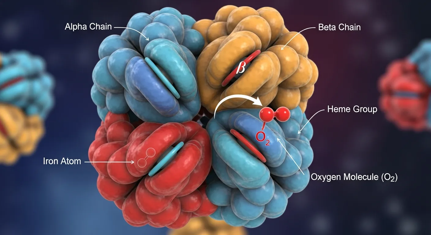
Hemoglobin structure and oxygen binding are essential for efficient oxygen transport throughout the body. Hemoglobin’s unique four-subunit structure allows it to bind and release oxygen as needed, ensuring tissues receive adequate oxygen for normal function. Understanding this process helps explain how blood carries oxygen and why hemoglobin disorders can affect overall health.
Hemoglobin structure and oxygen binding work together so red blood cells can carry oxygen. Hemoglobin has four globin chains and four heme groups. Each heme holds an iron atom that binds one oxygen molecule, and cooperative binding lets hemoglobin load and release oxygen efficiently.
Every breath you take depends on a single remarkable protein. Hemoglobin sits inside your red blood cells, picking up oxygen in your lungs and dropping it off where your body needs it most. Without it, oxygen could never travel far enough to keep your tissues alive.
What makes this protein so good at its job? The answer lies in its shape. The link between hemoglobin structure and oxygen binding is one of biology’s most elegant examples of form following function. Tiny changes in shape allow hemoglobin to grab oxygen tightly when it’s plentiful and let go just as readily when it’s scarce.
This guide breaks down hemoglobin structure and function in clear terms. You’ll learn how the molecule is built, how the oxygen binding mechanism of hemoglobin actually works, and why structural defects can lead to serious blood disorders like thalassemia and sickle cell disease. Whether you’re a student, a patient, a caregiver, or simply curious, you’ll come away with a solid understanding of how hemoglobin oxygen transport keeps you alive.
Why Is Hemoglobin So Important for Oxygen Transport?
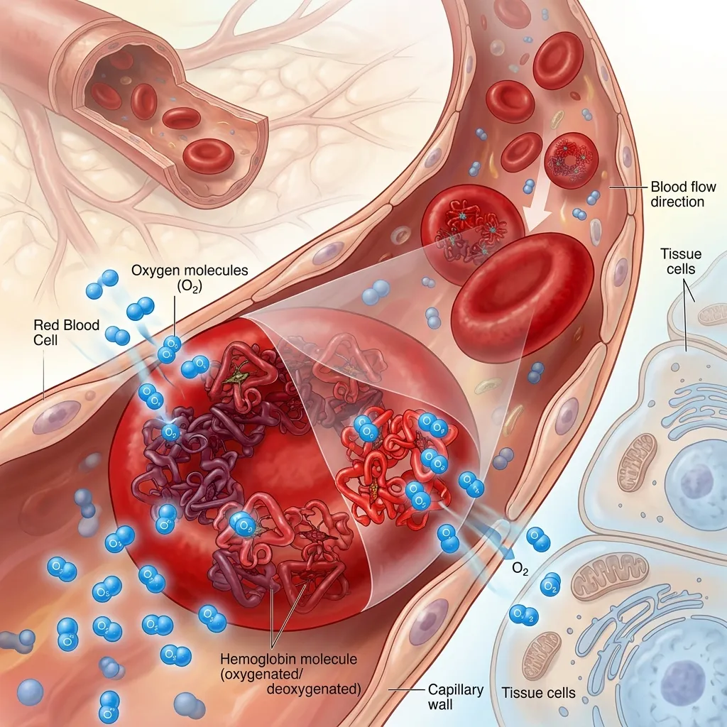 Hemoglobin is the protein responsible for carrying oxygen through your bloodstream. Each red blood cell holds roughly 270 million hemoglobin molecules, and each one can transport up to four oxygen molecules at a time. This massive carrying capacity is what makes hemoglobin oxygen transport so effective.
The role of hemoglobin goes beyond simply moving oxygen. It also helps carry carbon dioxide back to the lungs and plays a part in regulating blood pH. Red blood cells survive about 120 days in circulation, delivering oxygen to every tissue and organ throughout that lifespan.
When hemoglobin works properly, your cells receive the oxygen they need to produce energy. When hemoglobin structure and function break down—through genetic mutations or other defects—oxygen delivery suffers, and conditions like chronic anemia can develop.
What Is the Structure of Hemoglobin?
![Hemoglobin structure diagram showing four globin chains and heme groups]
Hemoglobin structure and oxygen binding cannot be understood without first looking at how the molecule is built. Like all proteins, hemoglobin has four levels of structural organization.
Primary, Secondary, Tertiary, and Quaternary Structure
The primary structure is the exact sequence of amino acids that make up each globin chain. Even a single error in this sequence can change how the whole protein behaves, as seen in sickle cell disease.
The secondary structure refers to how the chain folds into local shapes. Globin chains are rich in alpha-helices, twisting segments that give the protein stability.
The tertiary structure is the complete three-dimensional fold of a single globin chain. This fold creates a pocket that cradles the heme group and protects the iron atom inside.
The quaternary structure is the assembly of all four globin chains into a single functional unit. This four-part arrangement is essential because it allows the chains to communicate during oxygen binding.
The Heme Group: The Heart of Oxygen Binding
At the center of the oxygen binding mechanism of hemoglobin sits the heme group. Each hemoglobin molecule contains four heme groups, one tucked inside each globin chain.
Every heme group holds a single iron atom at its core. This iron atom is the exact spot where oxygen attaches. Because each hemoglobin molecule has four heme groups, it can bind four oxygen molecules at once. The iron must remain in its ferrous (Fe²⁺) state to bind oxygen correctly—if it oxidizes, it can no longer carry oxygen.
Globin Chains: Alpha, Beta, Gamma, and Delta
The globin portion of hemoglobin is made of four protein chains. The exact chains used depend on a person’s stage of life.
- Alpha chains: Found in nearly all forms of hemoglobin, produced by genes on chromosome 16.
- Beta chains: The main partner chains in adult hemoglobin (HbA), produced by the HBB gene on chromosome 11.
- Gamma chains: Found in fetal hemoglobin (HbF), which binds oxygen more tightly to pull it across the placenta.
- Delta chains: Found in the minor adult hemoglobin HbA2, usually less than 3.5 percent of total hemoglobin.
Normal adult hemoglobin (HbA) consists of two alpha chains and two beta chains. These chains must be produced in balanced amounts. When that balance breaks—as in thalassemia—red blood cells suffer. You can read more about this in our guide on alpha and beta globin chain imbalance.
Cooperative Binding and Allosteric Effects
Here’s where hemoglobin gets clever. When one oxygen molecule binds to a heme group, it slightly changes the shape of the whole protein. This change makes it easier for the next oxygen molecule to bind. This phenomenon is called cooperative binding.
Cooperative binding is an allosteric effect, meaning activity at one site affects activity at another. It’s the reason hemoglobin can load up on oxygen quickly in the lungs and release it efficiently in the tissues.
How Does the Oxygen Binding Mechanism of Hemoglobin Work?
![Oxygen molecule binding to heme iron within hemoglobin]
The oxygen binding mechanism of hemoglobin is a precise dance between chemistry and shape. Understanding it reveals why hemoglobin is so much better at its job than a simple oxygen sponge would be.
How Oxygen Binds to the Heme Iron
Oxygen binds directly to the iron atom at the center of each heme group. When an oxygen molecule attaches, it pulls the iron atom slightly into the plane of the heme ring. This tiny movement tugs on the globin chain attached to the iron, setting off a chain reaction throughout the protein.
Conformational Changes During Oxygen Binding
That small tug does something powerful. It shifts the position of the other globin chains, gently nudging their heme groups into a shape that binds oxygen more easily. As more oxygen binds, the protein becomes progressively better at grabbing even more. This is the molecular basis of cooperative binding.
The Tense (T) and Relaxed (R) States
Scientists describe hemoglobin as existing in two main shapes. The Tense (T) state has a low affinity for oxygen and dominates when oxygen levels are low, such as in the tissues. The Relaxed (R) state has a high affinity for oxygen and dominates when oxygen is plentiful, such as in the lungs.
As oxygen binds, hemoglobin shifts from the T state toward the R state. As oxygen is released, it shifts back. This back-and-forth is what makes hemoglobin oxygen transport so responsive to the body’s needs.
Factors That Affect Oxygen Affinity: The Bohr Effect and 2,3-BPG
Several factors fine-tune how tightly hemoglobin holds oxygen.
The Bohr effect describes how lower pH (more acidic conditions) and higher carbon dioxide reduce hemoglobin’s affinity for oxygen. Active tissues produce more carbon dioxide and acid, so hemoglobin releases more oxygen exactly where it’s needed most.
2,3-bisphosphoglycerate (2,3-BPG) is a molecule inside red blood cells that lowers oxygen affinity. It helps hemoglobin release oxygen to the tissues. Levels of 2,3-BPG rise at high altitudes, helping the body adapt to thinner air.
How Does Hemoglobin Transport Oxygen Through the Body?
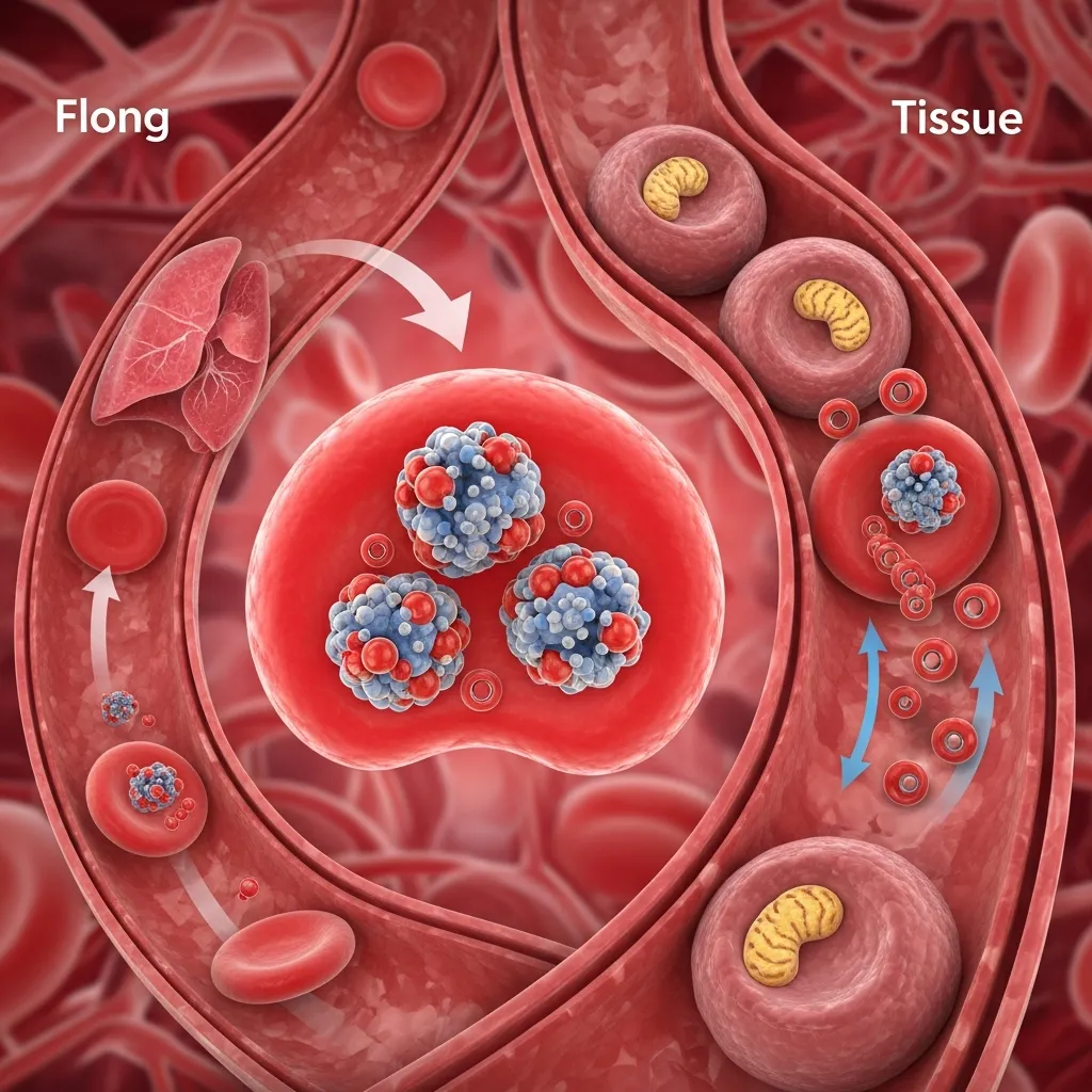 The link between hemoglobin structure and function comes alive when you follow oxygen on its journey from lungs to tissues and back again.
Oxygen Uptake in the Lungs
In the lungs, oxygen levels are high. Hemoglobin shifts into its high-affinity R state and rapidly binds oxygen. By the time blood leaves the lungs, hemoglobin is almost fully saturated—around 97 to 98 percent loaded with oxygen.
Oxygen Release in the Tissues
In the tissues, oxygen levels are low and carbon dioxide is high. Thanks to the Bohr effect and the T state, hemoglobin releases its oxygen where cells need it. This targeted delivery is the whole point of hemoglobin oxygen transport.
Carbon Dioxide Transport and the Haldane Effect
Hemoglobin doesn’t just carry oxygen. It also helps transport carbon dioxide from the tissues back to the lungs. The Haldane effect describes how deoxygenated hemoglobin carries carbon dioxide more readily. When hemoglobin releases oxygen in the tissues, it picks up carbon dioxide more easily—and the reverse happens in the lungs.
The Role of Hemoglobin in pH Regulation
Hemoglobin acts as a buffer, helping keep blood pH stable. By binding hydrogen ions released during carbon dioxide transport, it prevents the blood from becoming too acidic. This buffering role is a quiet but vital part of hemoglobin function.
What Happens When Hemoglobin Structure Goes Wrong?
![Comparison of normal and abnormal hemoglobin affecting red blood cells]
When mutations alter hemoglobin structure and function, the consequences can be serious. These disorders show just how much oxygen transport depends on getting the structure exactly right.
Fetal Hemoglobin and Its Significance
Fetal hemoglobin (HbF) uses gamma chains instead of beta chains. This gives it a higher oxygen affinity, which lets the fetus pull oxygen from the mother’s blood. After birth, the body switches from HbF to adult HbA. This handoff is controlled by a precise process explained in our guide on the globin gene switching mechanism.
Hemoglobinopathies: Sickle Cell Anemia and Thalassemia
Sickle cell anemia results from a single mutation in the beta-globin chain. This change produces abnormal hemoglobin S, which causes red blood cells to distort into rigid sickle shapes when oxygen levels drop. These cells block blood vessels and cause pain.
Thalassemia results from reduced production of either alpha or beta globin chains. The imbalance leaves excess chains that damage developing red blood cells. This contributes to chronic anemia and other complications, often through a process called ineffective erythropoiesis.
How Structural Abnormalities Affect Oxygen Binding
Both sickle cell disease and thalassemia interfere with normal hemoglobin oxygen transport. In sickle cell disease, the abnormal shape reduces how well red blood cells carry and deliver oxygen. In thalassemia, fewer functional hemoglobin molecules mean less oxygen reaches the tissues. To understand the wider category of these conditions, see our guide on hemoglobin synthesis disorders.
What Are the Advanced Concepts in Hemoglobin Biology?
Hemoglobin remains a focus of cutting-edge research because so much medicine depends on understanding it.
Allosteric Regulation and Protein Dynamics
Hemoglobin is one of the most studied allosteric proteins in all of biology. Its ability to switch between the T and R states makes it a model system for understanding how proteins respond to their environment. Researchers use it to explore how shape and function are linked in living systems.
Computational Models of Oxygen Binding
Scientists now use computer simulations to model how oxygen binds to hemoglobin atom by atom. These computational models help predict how mutations change oxygen affinity and how new drugs might affect the protein. According to the Protein Data Bank, hemoglobin was among the first proteins ever mapped in three dimensions, which laid the groundwork for this kind of research.
Therapeutic Strategies Targeting Hemoglobin
Several treatments now target hemoglobin directly. Drugs like hydroxyurea boost fetal hemoglobin to ease sickle cell disease. Newer gene-editing approaches, including CRISPR-based therapies, aim to correct the underlying mutations. These strategies show how a deep understanding of hemoglobin structure and oxygen binding leads directly to better treatments.

Hemoglobin is a masterpiece of biological engineering. Its four-chain structure, four heme groups, and shifting shape allow it to load oxygen in the lungs and release it in the tissues with remarkable precision. The close relationship between hemoglobin structure and function is what makes efficient oxygen transport possible.
Understanding the oxygen-binding mechanism of hemoglobin matters far beyond the classroom. It explains how disorders like thalassemia and sickle cell disease develop, and it guides the design of new treatments that are already changing lives.
If you want to keep learning, explore our related guides on hemoglobin synthesis disorders and the globin gene switching mechanism. And if you or a family member may be affected by a hemoglobin disorder, speak with a hematology specialist about diagnosis and the latest treatment options.
Frequently Asked Questions
1. What is hemoglobin structure and oxygen binding?
Hemoglobin structure and oxygen binding describes how the protein is built and how it carries oxygen. Hemoglobin has four globin chains and four heme groups. Each heme holds an iron atom that binds one oxygen molecule, allowing each hemoglobin to carry four oxygen molecules.
2. How does hemoglobin bind oxygen?
Oxygen binds to the iron atom inside each heme group. When the first oxygen attaches, it changes the protein’s shape, making it easier for more oxygen to bind. This process is called cooperative binding.
3. What are the four globin chains in hemoglobin?
The four globin chain types are alpha, beta, gamma, and delta. Adult hemoglobin (HbA) uses two alpha and two beta chains. Fetal hemoglobin uses gamma chains, and HbA2 uses delta chains.
4. What is the difference between the T and R states of hemoglobin?
The Tense (T) state has low oxygen affinity and releases oxygen in the tissues. The Relaxed (R) state has high oxygen affinity and binds oxygen in the lungs. Hemoglobin shifts between these states as it loads and unloads oxygen.
5. What is the Bohr effect?
The Bohr effect describes how lower pH and higher carbon dioxide reduce hemoglobin’s affinity for oxygen. This helps hemoglobin release more oxygen in active tissues, where carbon dioxide and acid levels are higher.
6. How does hemoglobin transport carbon dioxide?
Hemoglobin carries carbon dioxide from the tissues back to the lungs. The Haldane effect means deoxygenated hemoglobin binds carbon dioxide more readily, which improves carbon dioxide removal.
7. Why does fetal hemoglobin bind oxygen more tightly?
Fetal hemoglobin (HbF) uses gamma chains, which give it a higher oxygen affinity than adult hemoglobin. This allows the fetus to pull oxygen across the placenta from the mother’s blood.
8. How do sickle cell disease and thalassemia affect oxygen binding?
In sickle cell disease, a mutation distorts red blood cells into rigid shapes that carry oxygen poorly. In thalassemia, reduced globin chain production leaves fewer functional hemoglobin molecules, lowering oxygen delivery to the tissues.
9. What is 2,3-BPG and what does it do?
2,3-bisphosphoglycerate (2,3-BPG) is a molecule in red blood cells that lowers hemoglobin’s oxygen affinity. It helps release oxygen to the tissues and rises at high altitudes to help the body adapt.
10. Can hemoglobin disorders be treated?
Yes. Treatments include blood transfusions, iron chelation, and drugs like hydroxyurea that boost fetal hemoglobin. Gene therapy and CRISPR-based editing are emerging options that target the underlying genetic cause.
