
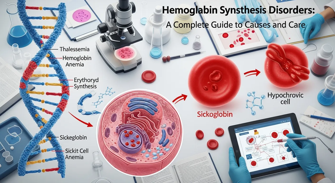
Hemoglobin synthesis disorders are inherited conditions that disrupt the body’s ability to make normal hemoglobin, the oxygen-carrying protein in red blood cells. The main types include thalassemia, sickle cell disease, and hemoglobin E disease. Early diagnosis, genetic counseling, and modern treatments help patients live healthier lives.
Hemoglobin synthesis disorders affect millions of people around the world, yet many remain undiagnosed for years. These genetic conditions disrupt how the body produces hemoglobin—the protein responsible for carrying oxygen from your lungs to every tissue. When that production breaks down, the result is anemia, fatigue, and a wide range of complications that vary from mild to life-threatening.
This guide explains what hemoglobin synthesis disorders are, how they develop at the genetic level, and the main categories you should know about. You’ll learn how doctors diagnose these conditions, what treatment options exist today, and what the future holds for patients and families. Whether you are a patient, a caregiver, or simply seeking clear answers, this resource breaks down complex science into practical, understandable information.
Understanding these disorders matters for one simple reason: knowledge changes outcomes. Carriers who know their status can plan families wisely. Patients who get diagnosed early can prevent organ damage. And families armed with accurate information can advocate for the care their loved ones deserve.
What Are Hemoglobin Synthesis Disorders?
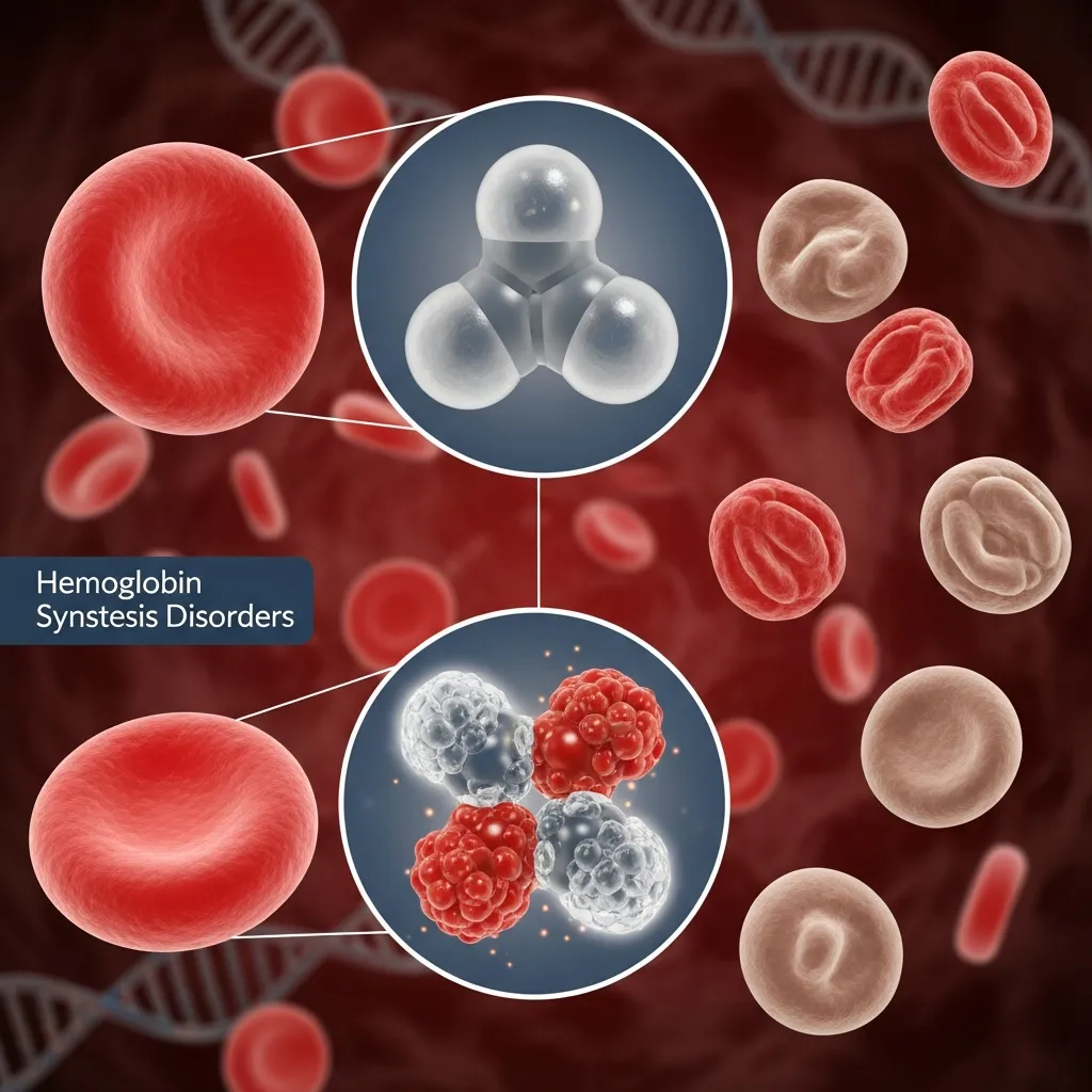 Hemoglobin synthesis disorders are a group of inherited blood conditions that interfere with the normal production of hemoglobin. Some texts refer to them as genetic hemoglobin disorders, disorders of hemoglobin production, or hemoglobin formation abnormalities. Whatever the label, the core problem is the same—the body cannot make enough normal, functional hemoglobin.
These disorders fall into two broad categories. The first involves reduced production of normal globin chains, which causes the thalassemia syndromes. The second involves the production of structurally abnormal hemoglobin, which causes conditions like sickle cell disease and hemoglobin E disease. Both categories trace back to mutations in the genes that direct hemoglobin assembly.
According to the World Health Organization (WHO, 2022), these inherited blood disorders are most prevalent in Mediterranean, Middle Eastern, South Asian, and Southeast Asian populations—regions where malaria has historically been common. Carriers may have gained a survival advantage against severe malaria, which helps explain why these mutations persist.
How Does the Body Make Hemoglobin?
To understand what goes wrong in hemoglobin synthesis disorders, it helps to know how hemoglobin is built in the first place.
The Molecular Structure of Hemoglobin
Hemoglobin is a complex protein made of two key components: heme and globin. The heme group contains an iron atom at its center, and this iron is what actually binds oxygen. Each hemoglobin molecule carries four heme groups, allowing it to transport four oxygen molecules at once.
The globin portion consists of four protein chains arranged in pairs. Normal adult hemoglobin (HbA) is made of two alpha-globin chains and two beta-globin chains. The body also produces other globin chains—gamma chains form fetal hemoglobin (HbF) before birth, while delta chains form a minor adult hemoglobin called HbA2. These chains must be produced in precise balance for hemoglobin to function correctly.
How Genes Control Globin Chain Production
The instructions for making globin chains come from specific genes. Alpha-globin production is controlled by genes on chromosome 16, while beta-globin production depends on the HBB gene on chromosome 11. When these genes carry mutations, the body either produces too little of a globin chain or produces a defective version. This imbalance is the root cause of every hemoglobin synthesis disorder.
Why Iron Matters in Hemoglobin Synthesis
Iron sits at the heart of every heme group, making it essential for oxygen transport. However, iron plays a complicated role in these disorders. Many patients absorb excess iron from their diet, and those who receive regular blood transfusions accumulate even more. Because the human body has no natural way to remove excess iron, it builds up in the heart, liver, and endocrine glands—where it can cause fatal organ damage if left untreated.
What Are the Major Categories of Hemoglobin Synthesis Disorders?
Genetic hemoglobin disorders span a wide spectrum, but most fall into a few well-defined groups.
Thalassemia Syndromes
Thalassemia is the umbrella term for disorders caused by reduced production of globin chains. The two main types are named for which chain is affected.
Alpha-Thalassemia
Alpha-thalassemia results from deletions or mutations in the alpha-globin genes. Because most people have four alpha-globin genes, the severity depends on how many are affected. Losing one or two genes usually causes mild or no symptoms. Losing three genes causes hemoglobin H disease, which leads to moderate to severe anemia. Losing all four genes causes Hb Barts hydrops fetalis, a condition that is typically fatal before or shortly after birth.
Diagnosis relies on blood tests and genetic analysis, while management ranges from no treatment for mild cases to regular transfusions for severe forms.
Beta-Thalassemia
Beta-thalassemia is caused by point mutations in the HBB gene on chromosome 11, which reduce or eliminate beta-globin production. More than 300 mutations have been identified. The condition is classified into three forms based on severity. Beta-thalassemia major (Cooley’s anemia) requires lifelong blood transfusions. Beta-thalassemia intermedia causes moderate anemia. Beta-thalassemia minor, or trait, usually causes minimal or no symptoms.
Symptoms of severe beta-thalassemia include chronic fatigue, pale skin, bone deformities, and an enlarged spleen. Treatment strategies center on regular red blood cell transfusions paired with iron chelation therapy to prevent iron overload. To explore the genetic roots of this condition in depth, see this detailed guide on beta thalassemia causes. If you want to understand carrier status and family planning, this beta thalassemia trait carrier guide offers clear, practical information.
Sickle Cell Disease
Sickle cell disease is one of the most well-known genetic hemoglobin disorders. It results from a single mutation in the HBB gene that produces an abnormal hemoglobin called hemoglobin S (HbS).
When oxygen levels drop, HbS causes red blood cells to change from their normal round shape into a rigid, crescent or “sickle” shape. These sickled cells are sticky and inflexible. They clump together and block small blood vessels, a process that triggers painful vaso-occlusive crises and damages organs over time. Patients may also experience acute chest syndrome, a serious lung complication, along with chronic anemia and increased infection risk.
Diagnosis uses hemoglobin electrophoresis and genetic testing. Management has advanced considerably—hydroxyurea reduces the frequency of pain crises, while newer gene therapy approaches now offer the potential for a functional cure.
What Other Genetic Hemoglobin Disorders Should You Know?
 Beyond thalassemia and sickle cell disease, several other hemoglobin formation abnormalities deserve attention.
Hemoglobin E Disease
Hemoglobin E disease is caused by a specific mutation in the HBB gene known as HBB:c.79G>A, which changes one amino acid in the beta-globin chain. This condition is especially common in Southeast Asia, where carrier rates in some populations reach up to 60 percent.
On its own, hemoglobin E disease is usually mild, causing only minor anemia. The picture changes dramatically when a person inherits a hemoglobin E gene from one parent and a beta-thalassemia gene from the other. This compound condition, called hemoglobin E/beta-thalassemia, can range from moderate anemia to severe, transfusion-dependent disease. For a complete breakdown of this condition, read this guide to hemoglobin E disease.
Hereditary Persistence of Fetal Hemoglobin (HPFH)
Hereditary persistence of fetal hemoglobin (HPFH) is a benign condition in which the body continues producing fetal hemoglobin (HbF) into adulthood. Normally, HbF production switches off shortly after birth. In HPFH, genetic changes keep it active.
What makes HPFH remarkable is its protective effect. When HPFH occurs alongside sickle cell disease or beta-thalassemia, the continued production of fetal hemoglobin can significantly soften the severity of those conditions. This protective mechanism is exactly why drugs like hydroxyurea, which boost HbF production, work so well in treating sickle cell disease.
How Are Hemoglobin Synthesis Disorders Diagnosed?
Accurate diagnosis is the foundation of good care. Several tests work together to identify these disorders of hemoglobin production.
Laboratory Tests
The diagnostic process usually starts with a Complete Blood Count (CBC) and red blood cell indices. In most hemoglobin synthesis disorders, red blood cells appear small (low MCV) and pale (hypochromic). A key clue that distinguishes thalassemia trait from iron deficiency is a high or normal red blood cell count despite the small cell size.
The definitive tests are hemoglobin electrophoresis and High-Performance Liquid Chromatography (HPLC). These techniques separate and measure the different types of hemoglobin in the blood. An elevated HbA2 level above 3.5 percent, for example, is the hallmark of beta-thalassemia trait.
Finally, genetic testing through DNA analysis provides the most precise diagnosis. It identifies the exact mutation, predicts disease severity in offspring, and enables accurate prenatal testing.
Newborn Screening and Prenatal Diagnosis
Many countries now include hemoglobin disorders in newborn screening programs, allowing treatment to begin before complications develop. For couples who are both carriers, prenatal diagnosis offers another layer of information. Chorionic villus sampling (CVS) at 10 to 13 weeks and amniocentesis at 15 to 20 weeks can determine whether a fetus has inherited a serious hemoglobin disorder.
What Treatments Are Available for Hemoglobin Synthesis Disorders?
Treatment depends heavily on the specific disorder and its severity. For many patients, a combination of approaches works best.
Blood Transfusions and Iron Chelation Therapy
Regular red blood cell transfusions remain the cornerstone of treatment for severe forms like beta-thalassemia major. Transfusions maintain healthy hemoglobin levels, suppress ineffective bone marrow activity, and support normal growth in children.
The catch is iron overload. Every transfusion adds iron the body cannot remove on its own. This is where iron chelation therapy becomes critical. Chelation medications bind to excess iron and help the body excrete it, preventing heart failure, liver cirrhosis, and hormonal problems.
Bone Marrow Transplantation and Gene Therapy
Currently, a bone marrow or stem cell transplant is the only established cure for severe hemoglobin synthesis disorders. The procedure replaces defective bone marrow with healthy stem cells from a matched donor, usually a sibling. However, it carries significant risks and requires a perfect donor match.
Gene therapy represents the most exciting frontier. Approaches including lentiviral gene addition and CRISPR-based gene editing are already showing promising clinical results. These techniques correct or work around the faulty gene, offering the potential for a lasting cure without needing a donor.
Supportive Care and Emerging Therapies
Supportive care fills the gaps between major interventions. Folic acid supplements support red blood cell production, while a splenectomy may help patients whose enlarged spleen destroys too many blood cells. Patients with thalassemia minor should also be cautious with certain medications—a detailed list of thalassemia minor drugs to avoid can help carriers stay safe. Looking ahead, new drugs that help red blood cells mature properly are entering the market, potentially reducing transfusion needs for many patients.
What Is It Like Living With a Hemoglobin Synthesis Disorder?
 A diagnosis reshapes daily life, but it does not have to define it. Many patients with milder forms live completely normal lives, while those with severe forms can thrive with proper medical care.
Quality of life depends on consistent treatment and monitoring. Patients benefit from regular appointments to track hemoglobin levels, monitor iron accumulation through MRI scans, and protect heart and liver health. Catching complications early dramatically improves outcomes.
Genetic counseling plays a vital role for families. Couples who both carry a hemoglobin mutation face a 25 percent chance with each pregnancy of having a child with a severe disorder. Counseling provides clear, evidence-based information so families can make informed reproductive choices, including options like preimplantation genetic testing through IVF.
Finally, emotional support matters. Living with a chronic condition takes a toll, and patient advocacy groups, support communities, and mental health resources help patients and families carry that weight together.
Conclusion
Hemoglobin synthesis disorders are complex, but they are increasingly manageable. From thalassemia and sickle cell disease to hemoglobin E and HPFH, each condition tells a story of how a small genetic change can ripple through the body—and how modern medicine continues to push back.
The path forward starts with three concrete steps. First, get tested if you have a family history or come from a high-prevalence region. Second, confirm any diagnosis with HPLC or genetic testing rather than relying on a basic blood count. Third, seek genetic counseling before planning a family if you and your partner are both carriers.
Research into gene therapy and personalized medicine is transforming what was once a devastating diagnosis into a condition with real hope for a cure. For trusted clinical information, the Centers for Disease Control and Prevention and the World Health Organization offer reliable, up-to-date resources. Early detection and proactive care remain your strongest tools for a healthier future.
Frequently Asked Questions
1. What are hemoglobin synthesis disorders?
Hemoglobin synthesis disorders are inherited conditions that disrupt the body’s ability to produce normal hemoglobin. They include thalassemia, sickle cell disease, and hemoglobin E disease, all of which can cause anemia and related complications.
2. What causes hemoglobin formation abnormalities?
These abnormalities are caused by mutations in the genes that direct globin chain production—primarily the alpha-globin genes on chromosome 16 and the HBB gene on chromosome 11. The mutations are inherited from one or both parents.
3. Are all disorders of hemoglobin production inherited?
Yes. Hemoglobin synthesis disorders are genetic and passed from parents to children. They are not contagious and cannot be acquired through contact, diet, or environment.
4. What is the difference between thalassemia and sickle cell disease?
Thalassemia results from reduced production of normal globin chains, while sickle cell disease results from an abnormal hemoglobin (HbS) that distorts red blood cells into a sickle shape. Both are genetic hemoglobin disorders but involve different mechanisms.
5. How are hemoglobin synthesis disorders diagnosed?
Diagnosis involves a Complete Blood Count (CBC), hemoglobin electrophoresis or HPLC, and genetic testing. These tests identify abnormal hemoglobin types and pinpoint the exact mutation responsible.
6. Can hemoglobin synthesis disorders be cured?
A bone marrow or stem cell transplant is currently the only established cure for severe forms. Gene therapy is an emerging option showing strong clinical promise and may offer functional cures without a donor.
7. Who is most at risk for these disorders?
People of Mediterranean, Middle Eastern, South Asian, Southeast Asian, and Sub-Saharan African descent face a higher risk, as these mutations are more common in regions where malaria was historically widespread.
8. Do all patients need blood transfusions?
No. Many people with mild forms or carrier status need no transfusions at all. Only those with severe forms, such as beta-thalassemia major, require regular transfusions to maintain healthy hemoglobin levels.
9. Why is iron chelation therapy important?
Patients who receive frequent transfusions accumulate toxic levels of iron because the body cannot remove it naturally. Iron chelation therapy binds and removes this excess iron, preventing fatal damage to the heart and liver.
10. Should carriers seek genetic counseling before having children?
Yes. If both partners carry a hemoglobin mutation, each pregnancy carries a 25 percent chance of producing a child with a severe disorder. Genetic counseling helps couples understand their risks and explore family planning options.
